
A male patient, age 28, presented in 2014 with fractured anterior teeth 12-11-21 due to falling from bed at a hospital, while recovering from a heart transplant.
His medical doctors refused to any dental/surgical procedures so a non-ideal interim solution was selected. Seven years later, due to severe external root resorption of two roots, the decision was to extract teeth 12-11 and restore the area using a minimally invasive, two-guide technique.
This is a 4-year follow-up review of the case.
INTRODUCTION
Modern dentistry is guided by wellestablished biological, restorative, and esthetic principles; however, complex medical histories may require treatments that depart from ideal protocols.
In such cases, the clinical objective is to use patient- specific protocols that can deliver predictable outcomes, while using minimally invasive techniques which respect the systemic limitations.
MEDICAL BACKGROUND
The 28-year-old patient underwent heart transplantation in 2014 following severe heart failure, after a compatible donor was identified.
THE TRAUMA AND TREATMENT IN 2014
During the postoperative recovery period, the patient fell from his hospital bed and fractures of teeth 12, 11, and 21 (Figures 1 and 2).
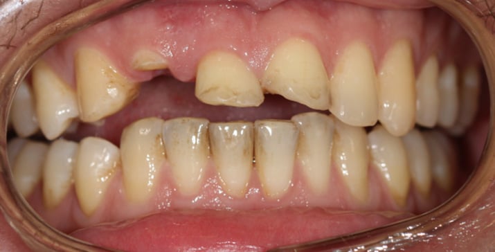
Figure 1. Pre-treatment view of teeth 12, 11, 21
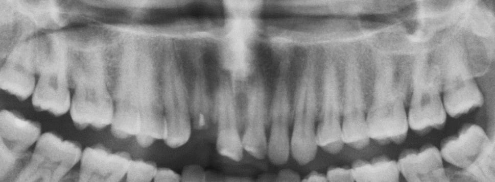
Figure 2. Pre-treatment radiograph
The treating medical team advised against any dental surgical intervention and requested that any dental treatment would be definitive, durable, and as conservative as possible.
Due of severe pain, root-canal treatment were performed, followed by a cast post-and-core and PFM crowns.
(Figures 3), knowing that this treatment was the only practical solution considering his medical condition.
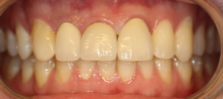
Figure 3. Post-treatment view of teeth 12, 11, 21 in 2014
THE TREATMENT IN 2021
A. REQUEST FOR A FIXED SOLUTION
Five years after the treatment of teeth 12, 11, and 21, the patient reported mild but persistent discomfort in the region of teeth 12 and 11. Periapical radiographs demonstrated severe external root resorption involving both teeth (Figure 4).
After consultation with the patient’s medical team, minimally invasive extraction of teeth 12 and 11 was performed under appropriate medical supervision. Given the extent of external resorption, immediate implant placement and immediate loading were ruled out.
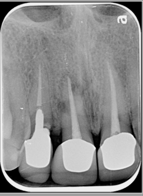
Figure 4. External root resorption.
Performed and documented by Dr. Nachum Samet Page 2 Three months after extraction, the patient requested a fixed restoration because he did not like the removable prosthesis as a long-term solution.
Based on CBCT imaging and intraoral scanning (Figures 5–7), the digital analysis approved the ability to use two narrow-platform (NP) 3.0×11.5mm Adin dental implants to replace the roots of 12 and 11 with optimal safety margins (Figure 8).
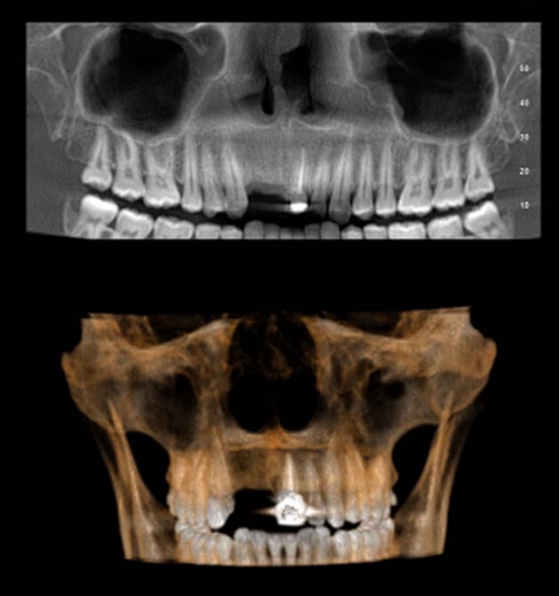
Figure 5. Pre-operative CBCT.
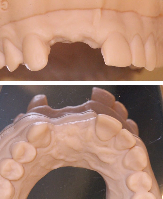
Figure 6. A printed model based on pre-operative intraoral scanning.
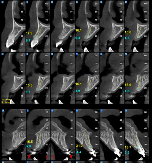
Figure 7. Pre-operative CBCT.
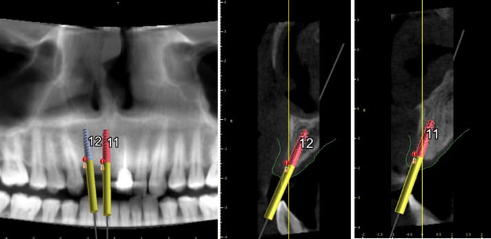
Figure 8. Digital plan for two NP implants.
SURGICAL AND RESTORATIVE GUIDES
Both a surgical guide and a restorative guide were designed and fabricated.
The surgical guide incorporated two near parallel 3.3mm NP sleeves (Figure 9) for use with the Adin' s NP Guided Surgery Kit .
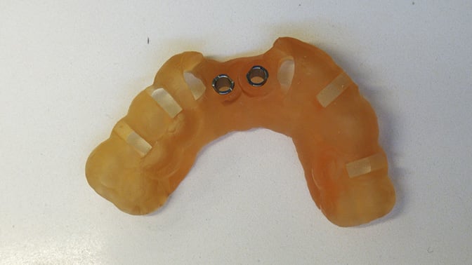
Figure 9. The surgical guide with two narrow sleeves.
To facilitate efficient provisional crown reconstruction, a restorative guide with hollow interim crowns was also fabricated (Figure 10 ).
The guide was made from white bis- acrylic material because alternative shades were not available at that time.
On the day of surgery, a minimally invasive protocol was followed in accordance the patient’s medical restrictions.
Correct seating of both surgical and restorative guides was verified first (Figure 11).
Local anesthesia was administered using 3% Mepivacaine.
A tissue punch was then used to remove a circular area of soft tissue at each implant site.
Initial osteotomy preparation was performed with a pilot drill to confirm accurate implant positioning (Figure 12), followed by drilling to the planned depth of 11.
5mm with the dedicated selfcentering drill (Figure 13).
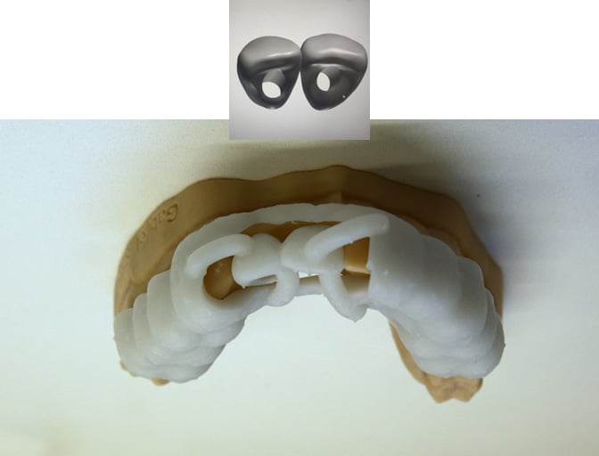
Figure 10. The Restorative guide placing the provisional crowns 11-12 in place.
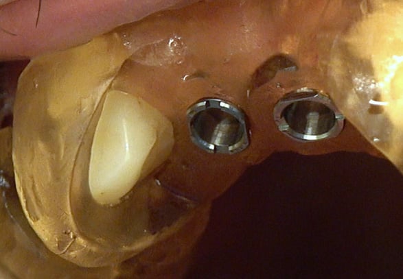
Figure 11. Verification that the surgical plan in accurately in place.
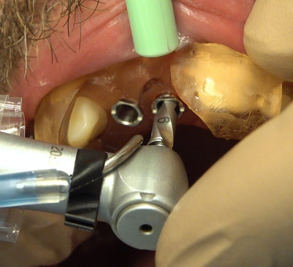
Figure 12. Initial drilling using the 6mm Pilot drill.
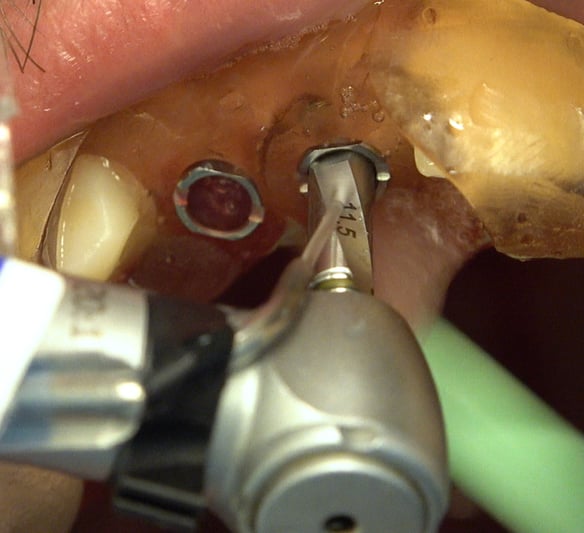
Figure 13. Drilling to the planned depth using 11.5mm drill.
Two implants were then placed according to the digital plan (Figures 14 and 15).
Once insertion torque of 60 Ncm was confirmed, immediate loading was performed.
Two Adin NP temporary cylinders were attached to the implants (Figure 16), and the restorative guide was seated to position the provisional crowns in place relative to the other teeth.
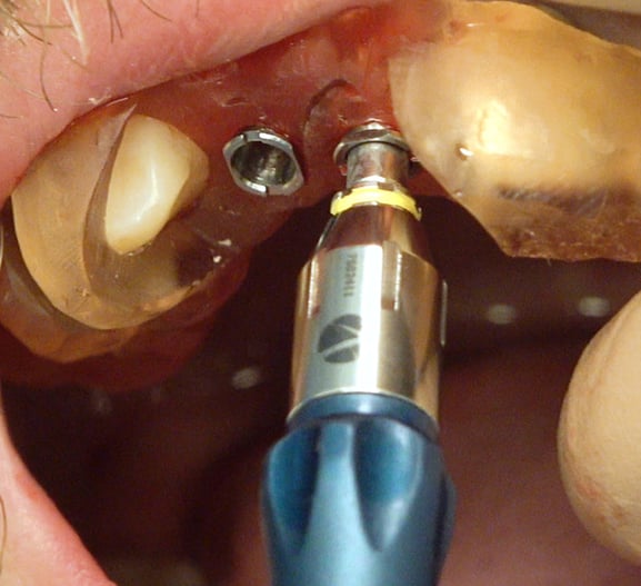
Figure 14. Through-the-guide implant placement.
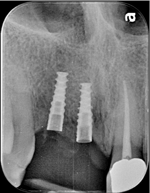
Figure 15. Implants in place
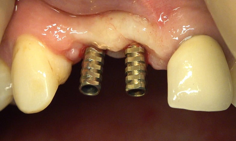
Figure 16. Adin’s NP interim abutments.
Using the brush technique, the provisional crowns were connected to the temporary abutments .
An ovate pontic shaped bases were created to support optimal emergence profile for each crown (Figures 17 and 18 ).
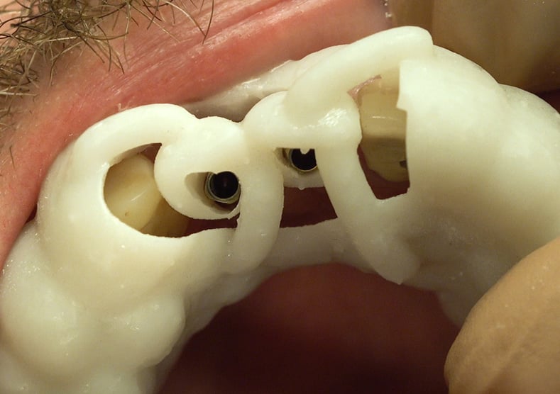
Figure 17. The restorative guide in place.
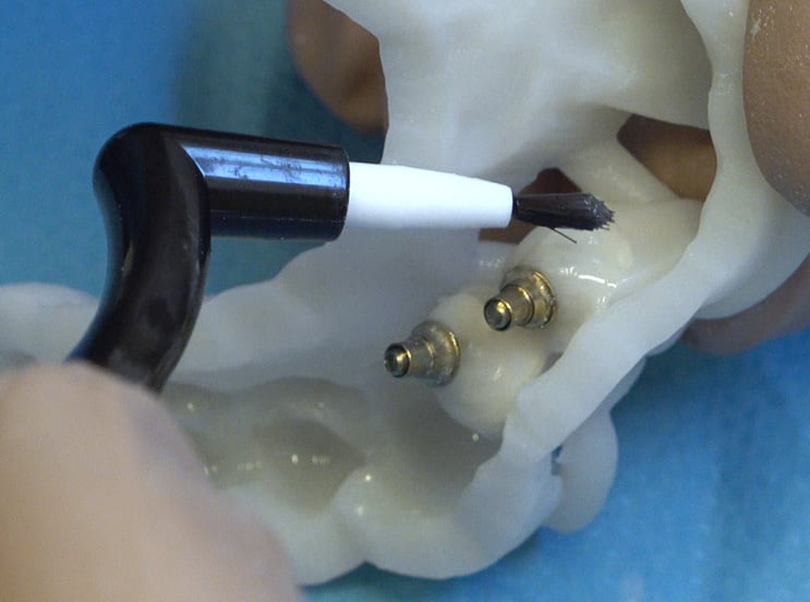
Figure 18. Brush technique used to finalize the emergence profile shape of the interim crowns.
Kerr acrylic tints were then used to alter the white shade of the material so the provisional crowns fit his natural teeth (Figures 19 and 20).
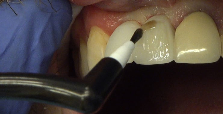
Figure 19. Altering the guide’s shade using Kerr acrylic tints.
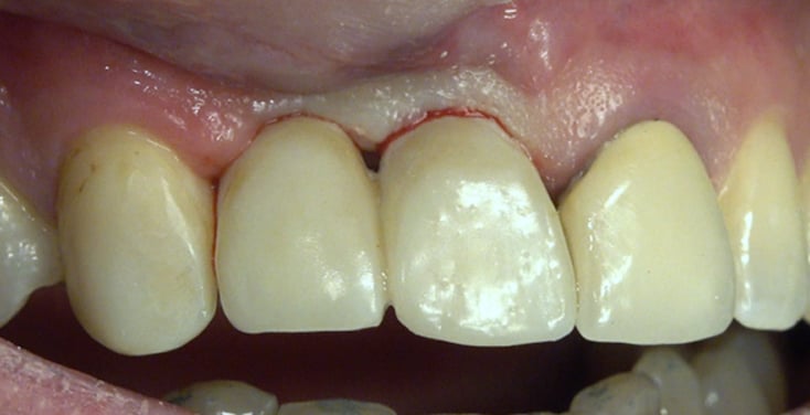
Figure 20. Interim crowns in place.
Eight weeks after surgery, Zirconia crowns with Ti- Base units were delivered.
Figure 21 presents the implants with the interim crowns 6 weeks after placement, and four years after placement. Figure 22 presents the restorative outcome four years after implant placement.
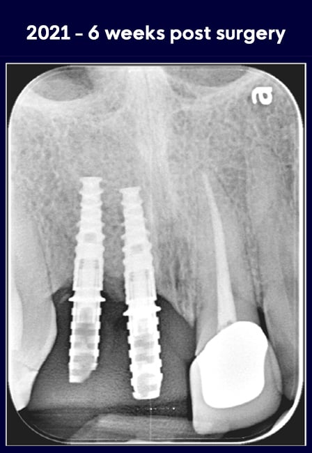
Figure 21. Radiographs presenting the interim and final crowns in place.
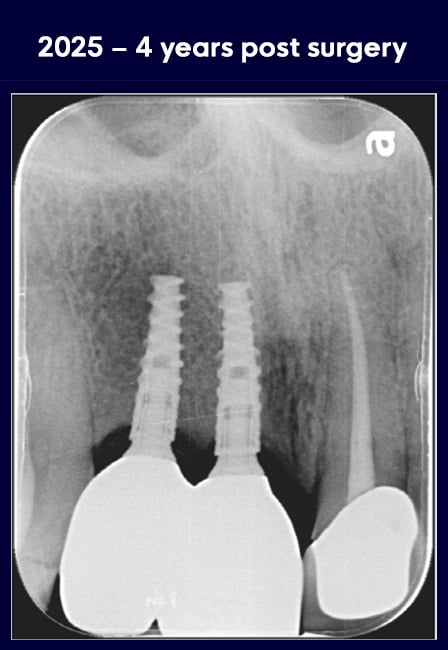
Figure 22. Presents the restorative outcome.
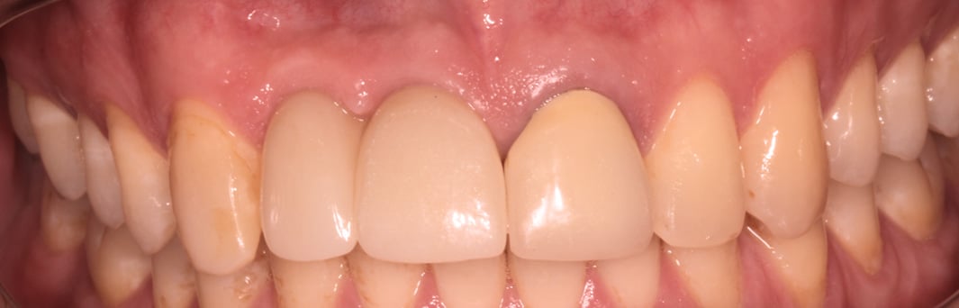
Figure 23. Final 12-11 crowns, 4 years after delivery.
CONCLUSIONS
Guided surgery offers significant advantages for both clinicians and patients and may be particularly valuable in medically complex cases where minimally invasive treatment is indicated.
In this case, the use of both a surgical guide and a restorative guide helped streamline treatment, reduce chair time, and support precise implant placement with immediate loading.
The dual-guide approach proved efficient and clinically effective in this medically complex setting and has since been adopted for the treatment of many other patients.



Leave a Comment